A Toronto Neurosurgeon Made a Radical Discovery About Brain Cancer

The brain surgeon Gelareh Zadeh discusses a new brain cancer therapy that could buy precious time for terminal patients
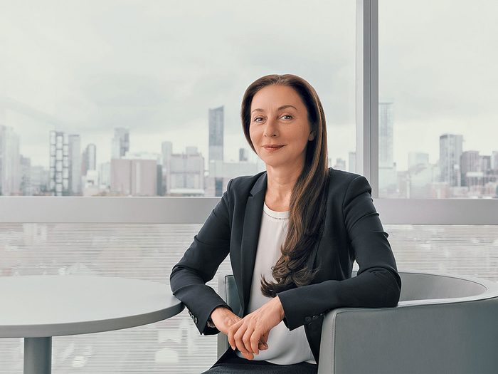
Before she pokes around in her patients’ brains, Gelareh Zadeh tries to put herself in their shoes. Brain cancer is a devastating disease: Glioblastoma, the most common and most lethal type of tumour, has an average survival time of just eight months, a length that hasn’t budged in decades. But delivering the news of this diagnosis isn’t so much a science as an art, one that Zadeh, a neurosurgeon at Toronto Western Hospital, and co-director at the Krembil Brain Institute, part of the University Health Network (UHN), has honed over 15 years in the field. Understanding who a patient is, how they were raised, whether they’re someone who prizes control—it all shapes the way she interacts with them. “How we manage the situation for patients impacts how they manage their disease,” she says. “We hold a unique position, because we’re engaged with them at the most sensitive time in their life.”
Zadeh, who left Iran as a teenager and landed in Winnipeg on a frigid Boxing Day in 1988, emphasizes that she can’t predict the future for any patient with brain cancer. But her research is, finally, moving the needle on the outcomes they can expect. Working with a colleague at the Princess Margaret Cancer Centre, she developed a blood test that can not only detect brain cancer but reveal the type of tumour and its likelihood to recur. And in a recent, groundbreaking clinical trial at the UHN, she helped identify a new combination therapy that may give patients with glioblastoma months or even years longer to live. Here, Zadeh discusses neurosurgery’s razor-thin margin of error, how to predict the risk for brain tumours and the value of staying in the moment.
What does the brain actually look like?
It’s incomparable. The layers that are there to protect our brain, the way the different structures of the brain reflect light—it’s just really beautiful and intricate. You can see the bundles of nerves that connect to each other and allow us to be who we are. I think that’s the part that really fascinates me. All of this intricate anatomy makes us uniquely who we are.
And what happens when something goes wrong in there?
We’re still in the infancy of understanding how we repair the brain. You can put stents in a heart, you can replace joints. But what’s the equivalent of a joint repair for the brain? Because once brain tissue is damaged—whether it’s through a stroke, neurodegeneration from Alzeheimer’s, aneurysm, brain cancer—the ability to restore that function is not there.
How is treating brain tumours different from treating other types of cancer?
Because of the eloquence of the brain, you have little margin of safety to reach a tumour. And it’s essential to remove the tumour without damaging brain tissue, because the likelihood of restoring that function is very low. That adds a degree of complexity and, I would say, stress to what we do. This is not to diminish what other surgeons do, but if you lose a few centimeters of your bowel during bowel surgery, the impact to the individual is not as tremendous. We have maybe a few millimeters that we can work in. The cranial nerves that allow us to talk, to move our eyes, to make facial expressions are so sensitive—in our world, we say that if you just look at the third nerve, it stops operating, because it’s such a sensitive nerve.
How has brain tumour diagnosis changed just in the time you’ve been in the field?
At the start of my career, we still relied on clinical exam to determine where the lesion was. Then magnetic resonance imaging came out, and it was one of the biggest evolutions in seeing, diagnosing and surgical planning. And on the research side, genomic analysis of tumours has really expanded our understanding of where these tumours come from and what are the potential targets.
What happens once we know the potential targets?
That provides us with a therapeutic approach. I also think we’re beginning to understand how we can use this data to come up with predictive modelling— meaning, what is the test that tells the average person whether they’re at risk? We all get a mammogram on a routine basis. There’s a PSA test for prostate cancer. So what is the single test that’s going to tell me I will be at risk of developing glioblastoma? What is the profile of my tumour that will distinguish whether I’ll respond to the standard treatment? Right now, we give radiation to everybody. And not everybody responds the same way.
Well, walk me through the blood test you’ve developed. What does it allow you to do?
The concern has always been that the blood-brain barrier, which protects [outside] material from going into our brain, also prevents us shedding material from the brain into the blood—like DNA evidence of cancer. In fact, we’ve demonstrated that, regardless of the type of brain tumour, it sheds pieces of DNA into the blood in a sufficient amount for us to study. We can detect brain cancer— and we can discriminate between the types of brain cancer, because you can have up to 150 types. And when you know what kind of tumour you’re dealing with, you can give the patient reassurance or give yourself direction. Then there’s the potential to use blood tests to tell when a tumour is coming back. Right now, we rely heavily on MRI, but MRI has limitations: You can only see pathologies that are bigger than, for example, 10 million cells, by which point it’s too late because the cancer’s already been quite active. So can a blood test tell us—faster, more accurately, earlier than an MRI—that cancer is coming back?
How likely is recurrence?
For glioblastoma, the likelihood of recurrence for a two-year period is 100 percent. It’s the most lethal adult cancer. The standard of treatment is surgery, followed by chemoradiation, but then, inevitably, recurrence. But we have a clinical trial that’s really remarkable. At recurrence, we inject an adenovirus [a weakened common-cold virus] into the tumour. The adenovirus is designed to attack cancer cells, but not normal brain tissue, and it’s delivered through a needle in a very slow, pressured process. After that injection, the patient goes on immunotherapy by oral intake of the drug. The adenovirus infects the cells and induces an immune reaction, and the immunotherapy comes in to really attack those cells and take away the dead cancer cells.
What results have you seen? It’s beyond exciting. For those who responded—who have signatures in their tumours that respond to this treatment— we have a 50 percent increase in survival. Some of our patients have lived for longer than three years. But also, I do want to encourage patients to focus on things outside of how long they have to live. You have to help people get to a place where they can enjoy the time they have.
Has this work made you more present as well? So much of our health can turn on a dime. I think the events that determine our lives and shape where we end up are moments we can’t actually predict or control. So I truly, firmly believe that I have to live in the moment.
Next: What Therapists Think About Neuro-Linguistic Programming for Mental Health


